Launch Offer
Valid until today,
The Easiest Way to Read an EKG Without Panicking!
The material for nurses, nursing students and new grads who need to recognize a rhythm, a block or an MI even when the cardiologist isn't there: explained with pictures, on real tracings.
I WANT TO READ EKGs WITH CONFIDENCESecure checkout · Instant download · 30-day guarantee
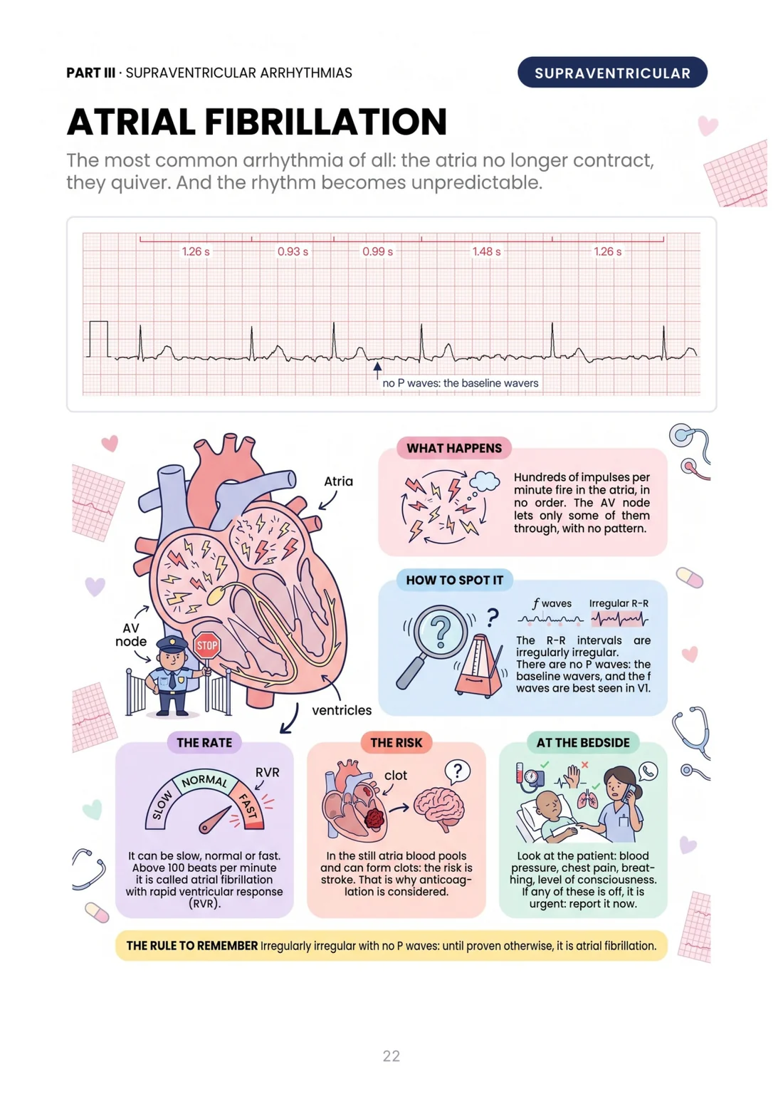
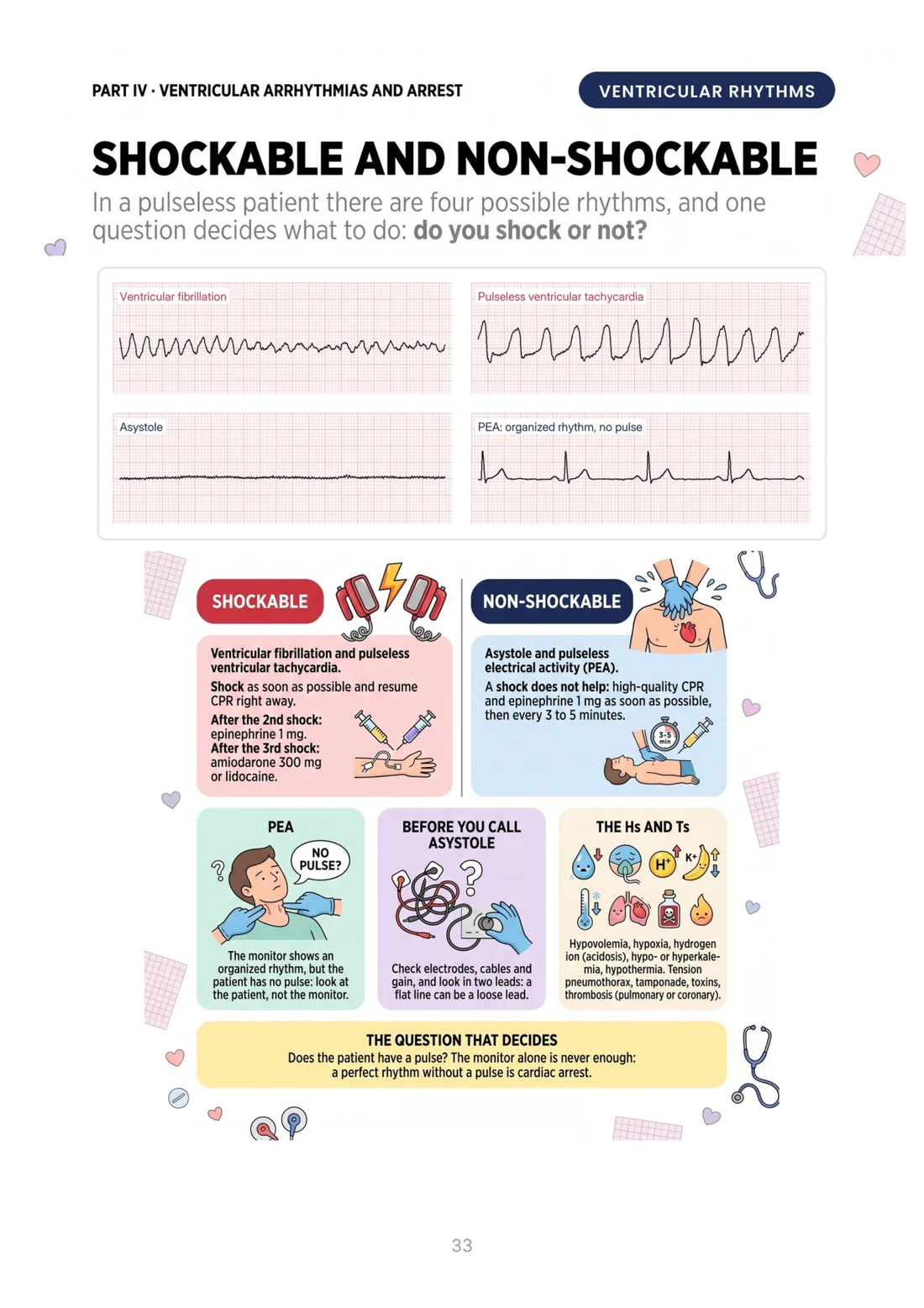
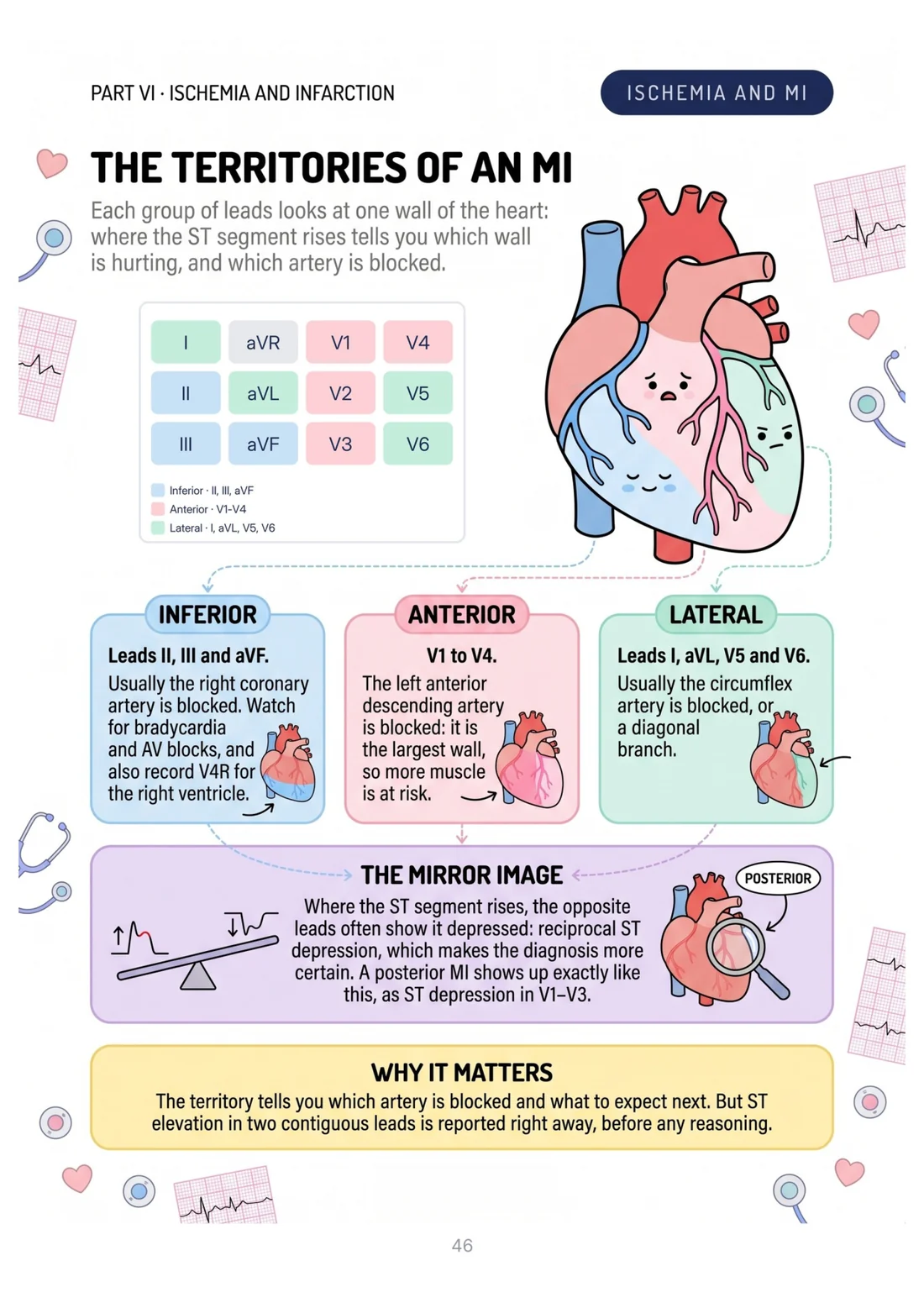
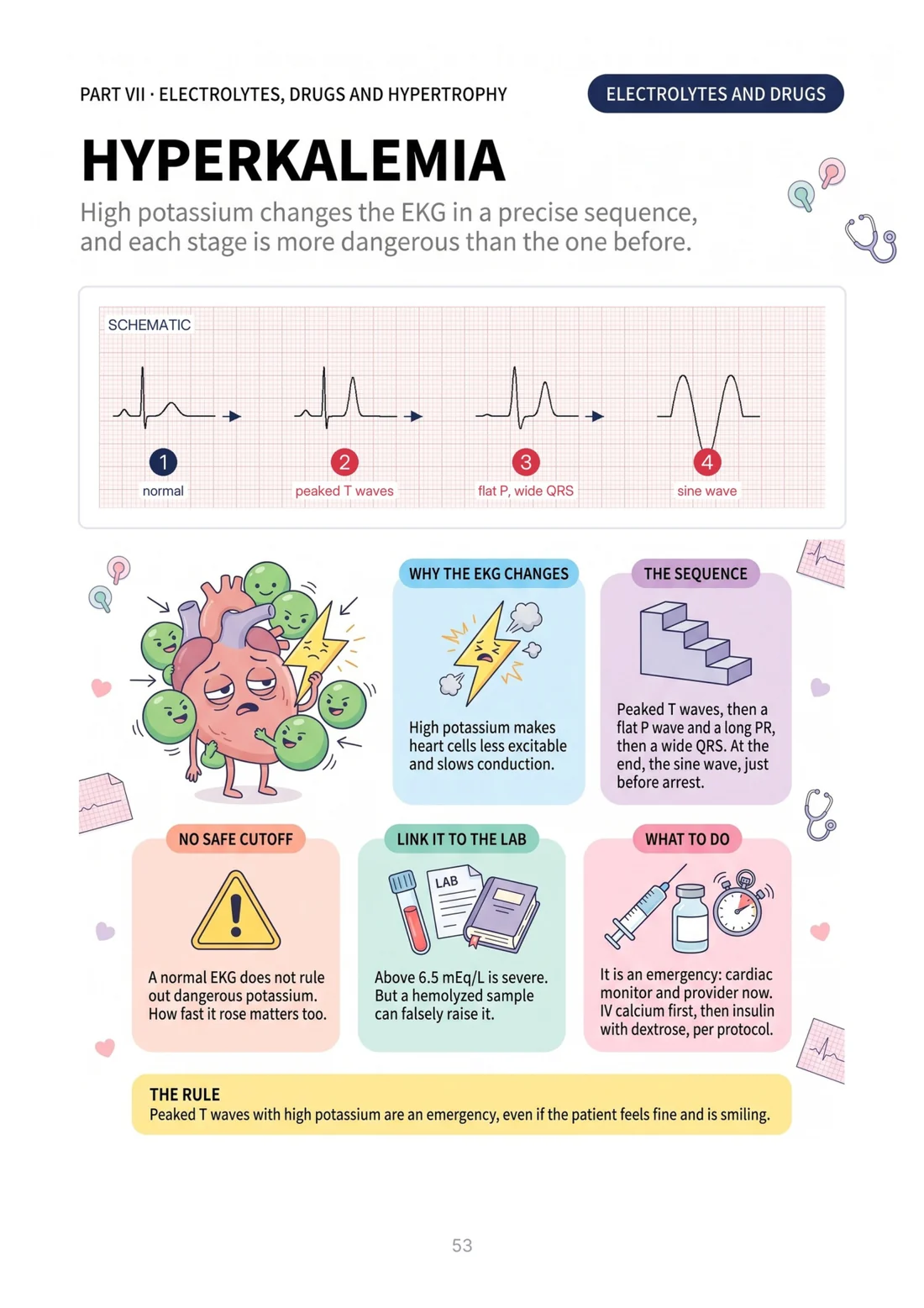
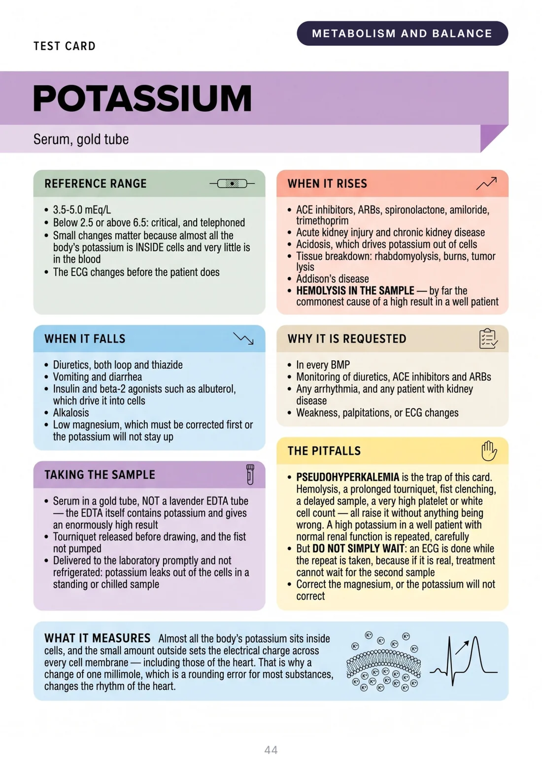
Here's What the Pages You'll Study Look Like
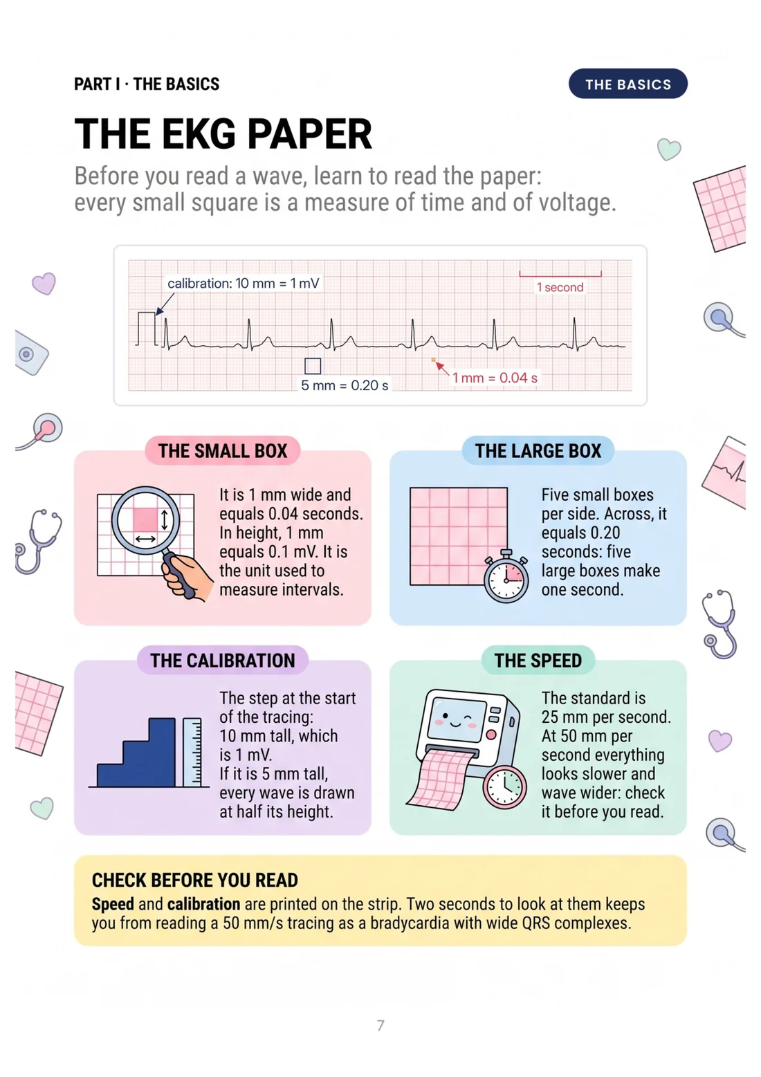
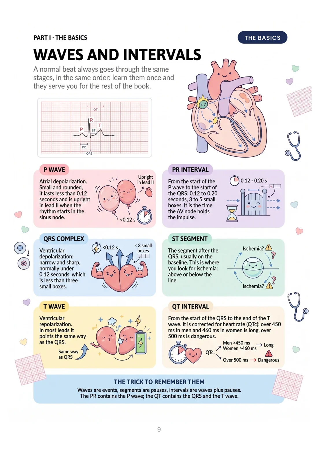
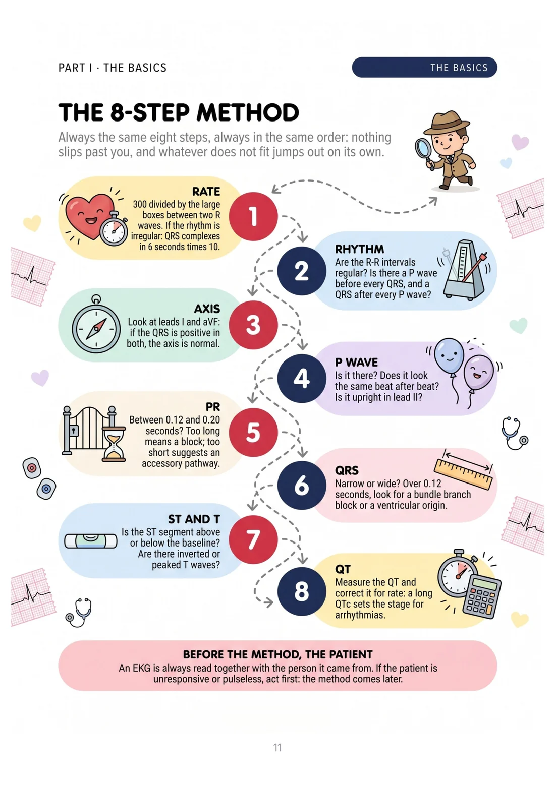
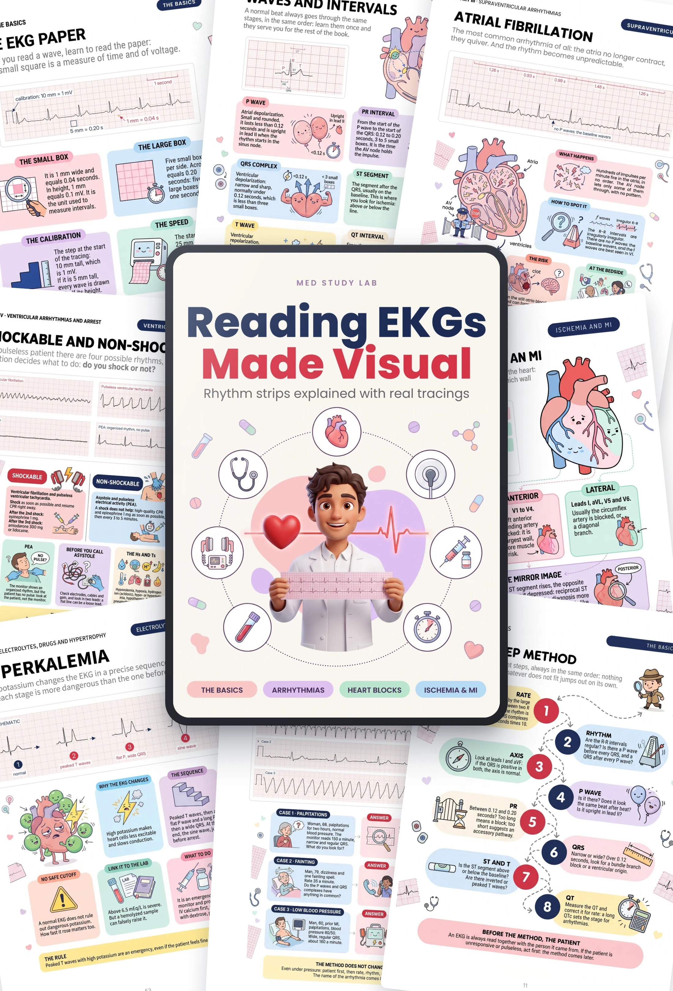
One topic per page, the real tracing in the middle and the drawing that tells you where to look. Look inside:
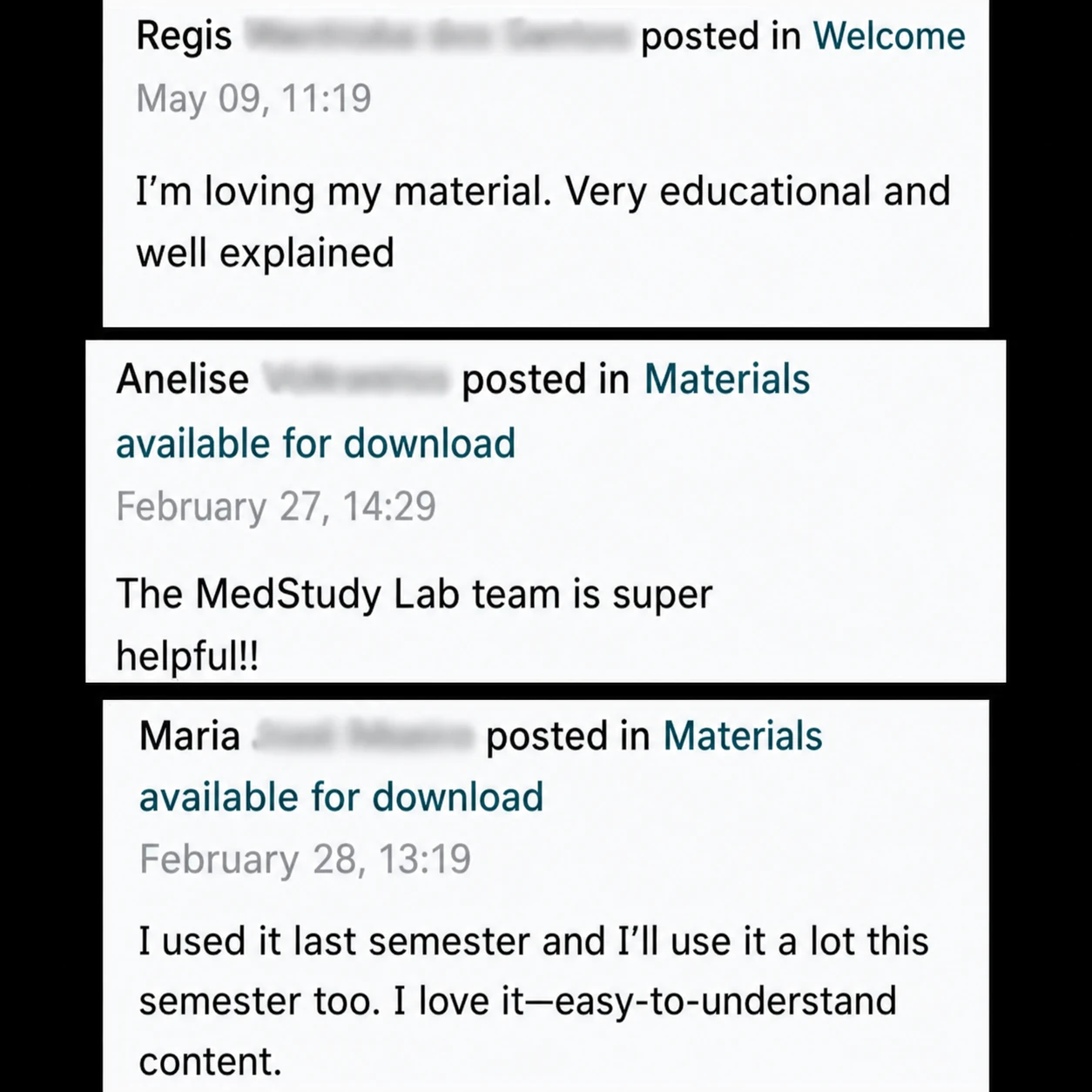
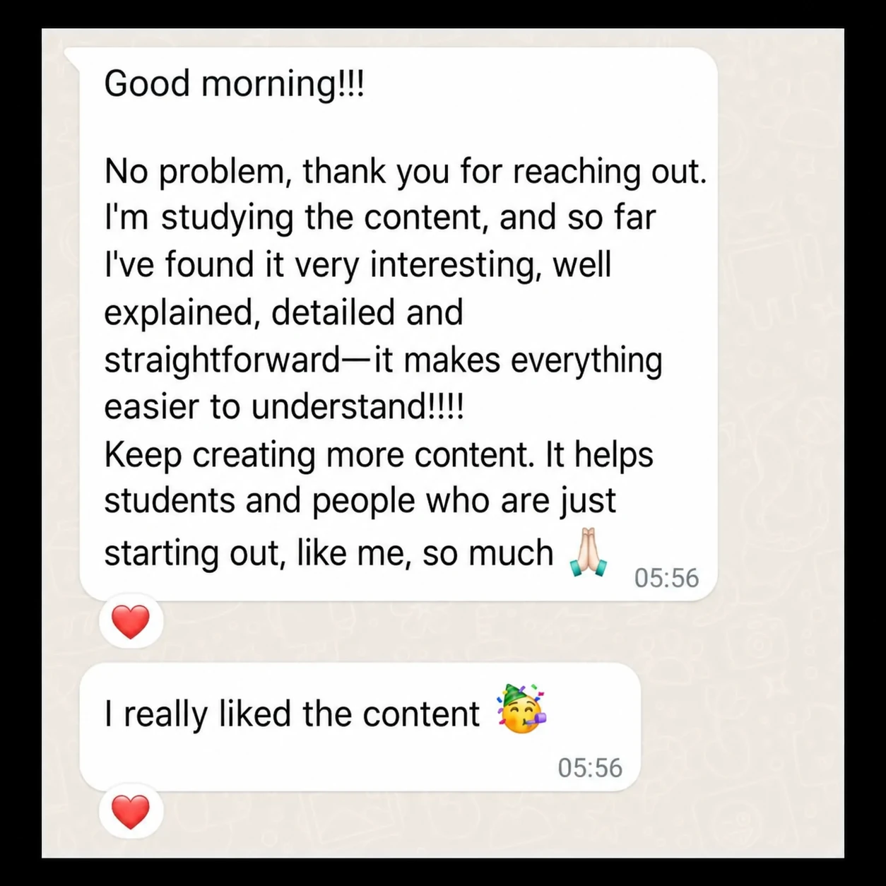
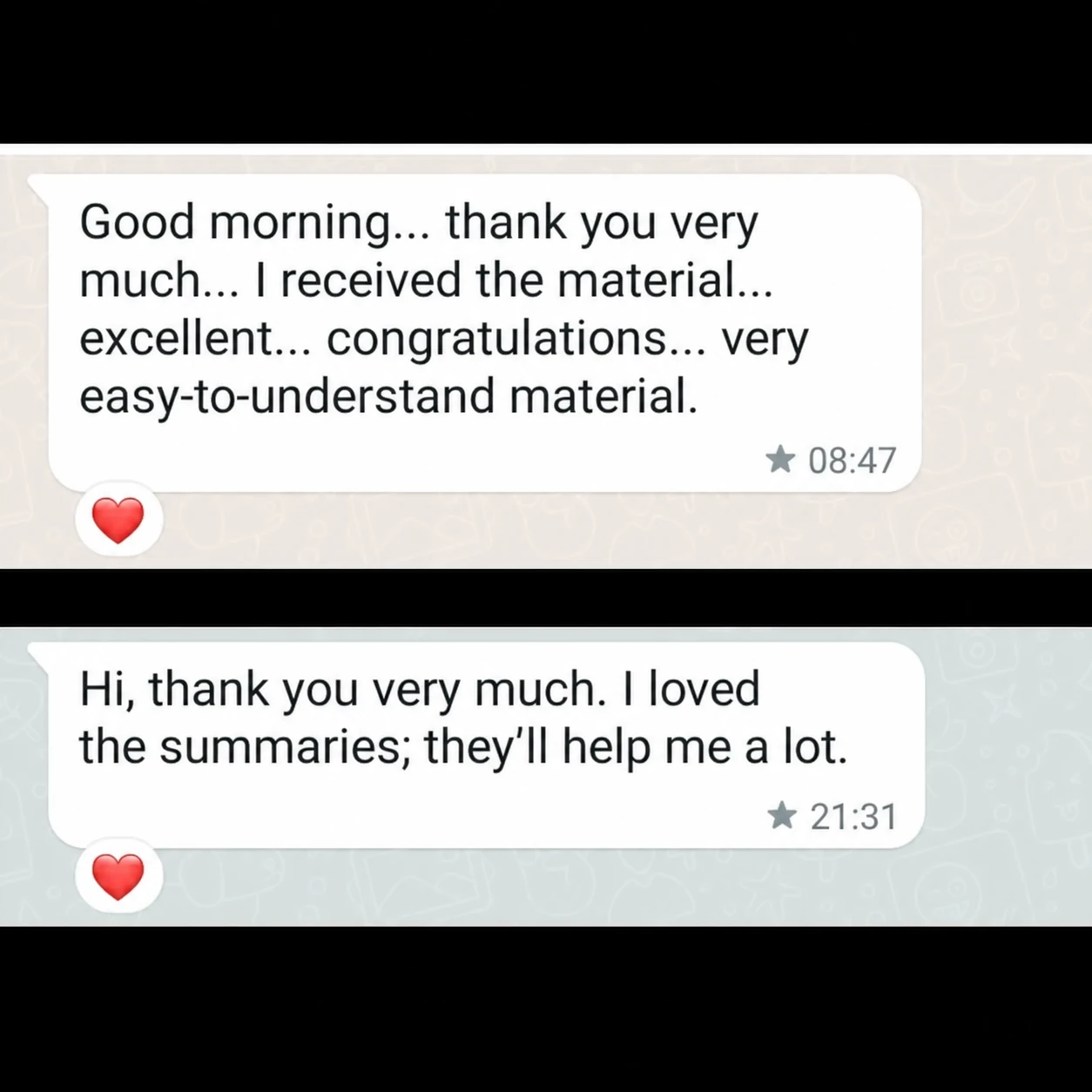
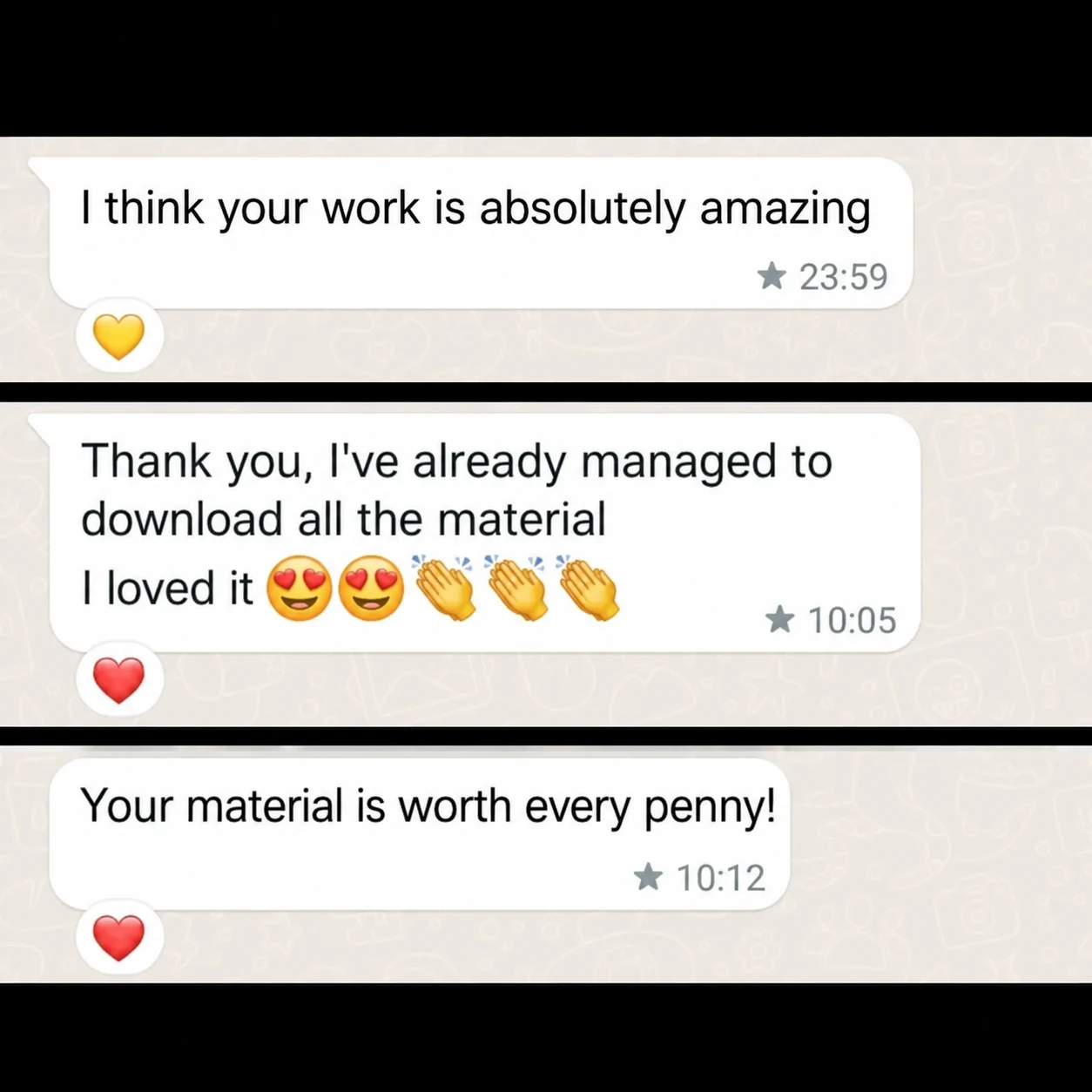
What Over 11,978+ Students Using MedStudyLab Have to Say:









Why the Tracings Are Real, Not Drawn
Almost every textbook shows perfect, hand-drawn waves: clean lines, no noise, every wave in its place.
Then you get to the unit and the strip in front of you looks nothing like the ones you studied.
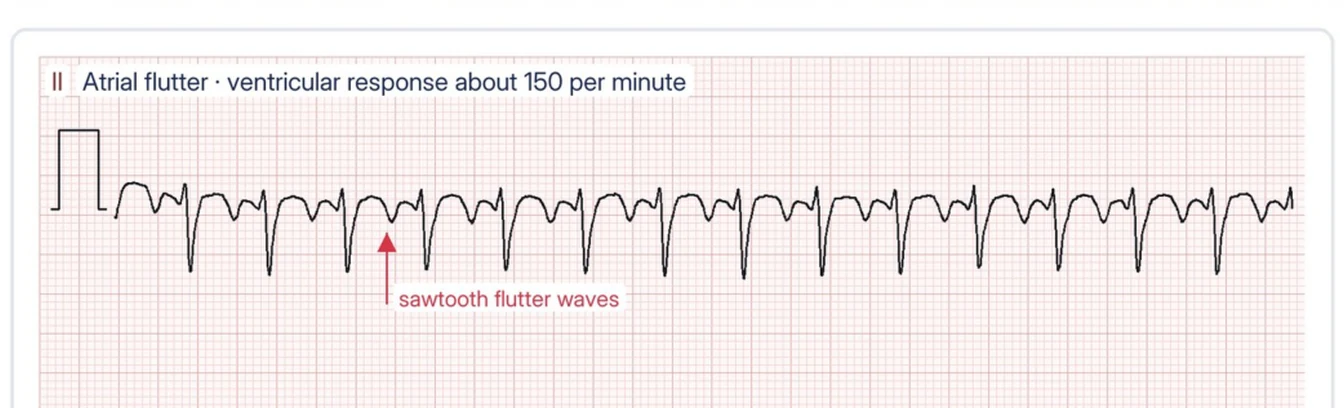
That's why every tracing in the book comes from PTB-XL: a public collection of 21,799 12-lead EKGs from 18,869 patients, each one read by cardiologists and published on PhysioNet.
They're printed on real EKG paper, at 25 mm per second and 10 mm per millivolt, just as they come out of the machine. The drawing around them tells you where to look; the tracing stays a real patient's.
So your eye trains on what you'll really see.
The Difference Is Visual
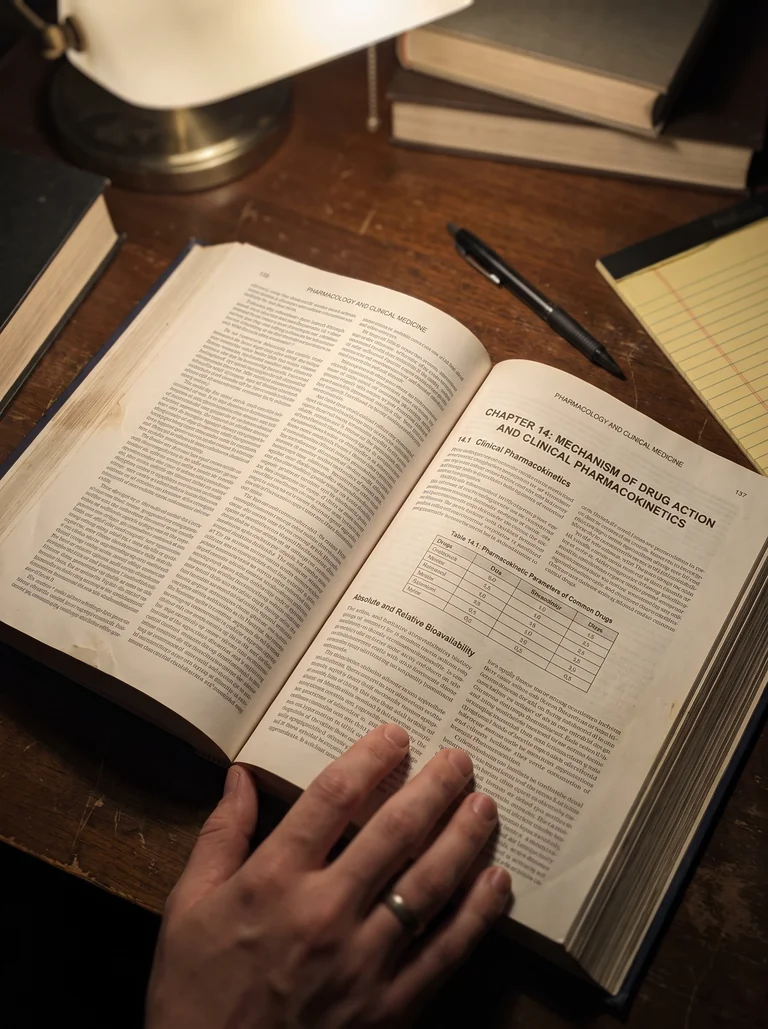
Stop staring at a tracing without knowing where to start:
FROM THIS
- Twelve leads and no order
- You don't know where to look
- The doubt comes when there's no time
TO THIS
- A method in eight steps
- The drawing shows you what to look for
- You recognize it at first glance
Table of Contents
Take a Look at the 8 Parts and 51 Chapters
From the EKG paper to MI, electrolytes and cases at the bedside: 68 pages, all illustrated
- The electrical heart
- The EKG paper
- The 12 leads
- Waves and intervals
- Where the electrodes go
- The 8-step method
- Part review
- Calculating the rate
- Normal sinus rhythm
- Sinus tachycardia and sinus bradycardia
- Sinus arrhythmia
- The axis in two leads
- Part review
- Premature atrial contractions
- Atrial fibrillation
- Atrial flutter
- Supraventricular tachycardia
- Wolff-Parkinson-White
- Part review
- Premature ventricular contractions
- Ventricular tachycardia
- Torsades de pointes
- Ventricular fibrillation
- Asystole and pulseless electrical activity
- Shockable and non-shockable
- Part review
- First-degree AV block
- Second-degree AV block
- Third-degree (complete) AV block
- Right bundle branch block
- Left bundle branch block
- Fascicular blocks
- Part review
- Ischemia, injury, infarction
- STEMI: the criteria
- The territories: inferior, anterior, lateral
- Posterior and right ventricle
- ST depression and NSTEMI
- Q waves and evolution
- STEMI mimics
- Part review
- Hyperkalemia
- Hypokalemia
- Calcium
- Long QT and drugs
- Digoxin
- Ventricular hypertrophy
- Part review
- Artifacts and reversed leads
- When to call right away
- Six guided cases
- Glossary — p. 65
- Sources and references — p. 67
- Index — p. 68
What If There Were an Easier Way?
Imagine this:
Instead of trying to remember fifty different patterns...
...you follow the same eight steps every time, in the same order: rate, rhythm, axis, P wave, PR, QRS, ST segment, QT.
Your brain stops searching at random. Every tracing becomes a checklist, and whatever doesn't fit jumps out on its own.
That's exactly what Reading EKGs Made Visual does.
It isn't “just another PDF”. It's a method in 8 parts and 51 illustrated chapters, built on real tracings, so you understand what you see instead of memorizing pictures.
Because when you know where to look, even the ugliest strip starts to make sense.
Here's What You Get With Reading EKGs Made Visual:
I
pp. 5–12
The Basics
The paper, the twelve leads and the waves: the vocabulary you need to read everything else.
II
pp. 13–19
Rate, Rhythm and Axis
Rate, rhythm and axis: the first three steps, done on every strip.
III
pp. 20–26
Supraventricular Arrhythmias
Atrial fibrillation, flutter and SVT: when the rhythm starts in the atria and races.
IV
pp. 27–34
Ventricular Arrhythmias and Arrest
From PVCs to the arrest rhythms, split into shockable and non-shockable.
V
pp. 35–42
Heart Blocks
AV blocks and bundle branch blocks: where the impulse slows down or stops.
VI
pp. 43–51
Ischemia and Infarction
The ST elevation you cannot miss, territory by territory, and what mimics it.
VII
pp. 52–59
Electrolytes, Drugs and Hypertrophy
Potassium, calcium and the drugs that prolong the QT: the EKG that changes with the labs.
VIII
pp. 60–64
At the Bedside
Artifacts and reversed leads, when to call right away, and six guided cases.
At the end: a glossary, sources and references and an index to find every topic.
But That's Not All: You Also Get 2 Bonus Books
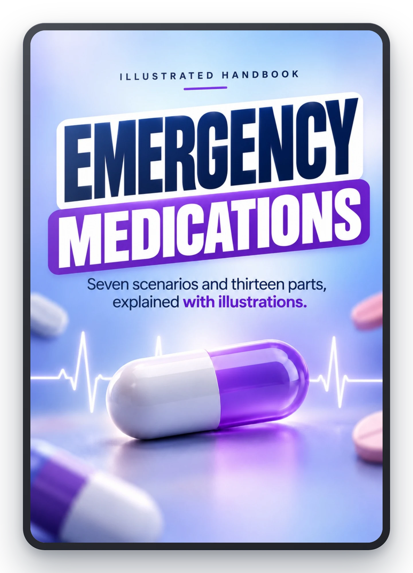
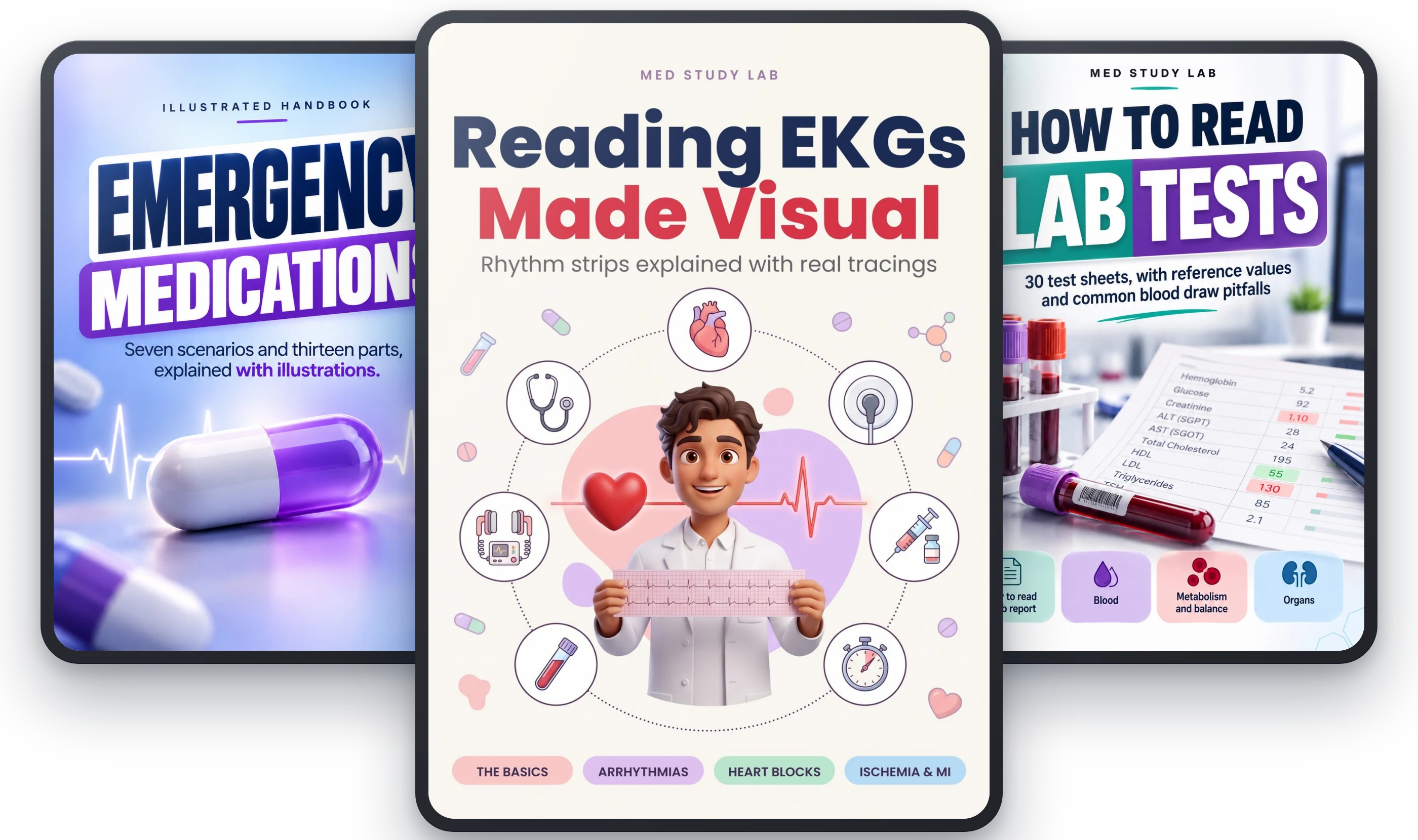
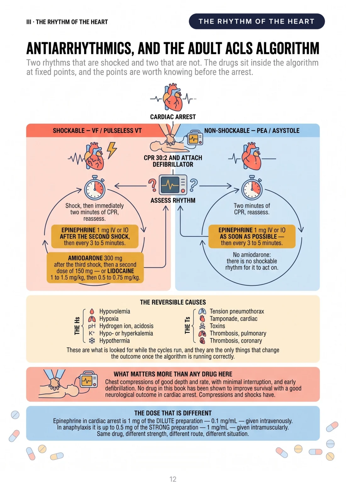
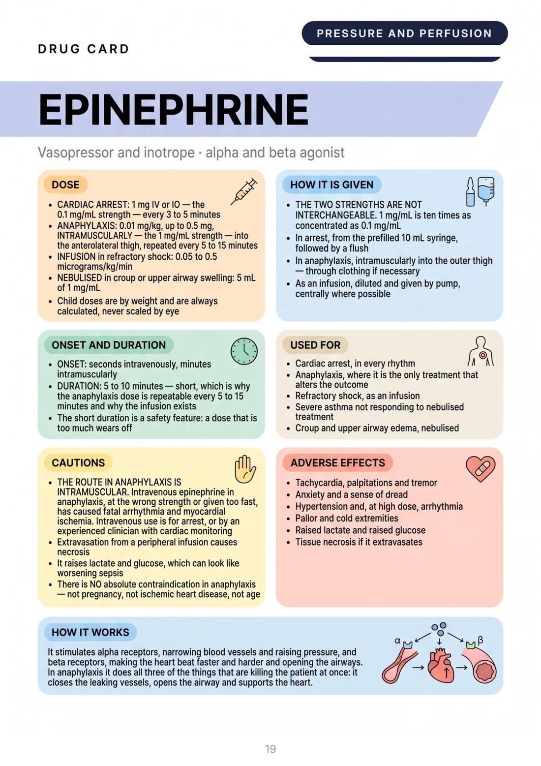
BONUS 1: “Emergency Medications”, the illustrated handbook
Standalone value: $19
82 pages on the drugs you use when a tracing becomes an emergency: adenosine, amiodarone, atropine and the rest of the crash cart, with the doses and dilutions to know by heart.
It completes Part IV of the book: you recognize the rhythm, and you already know what gets prepared.
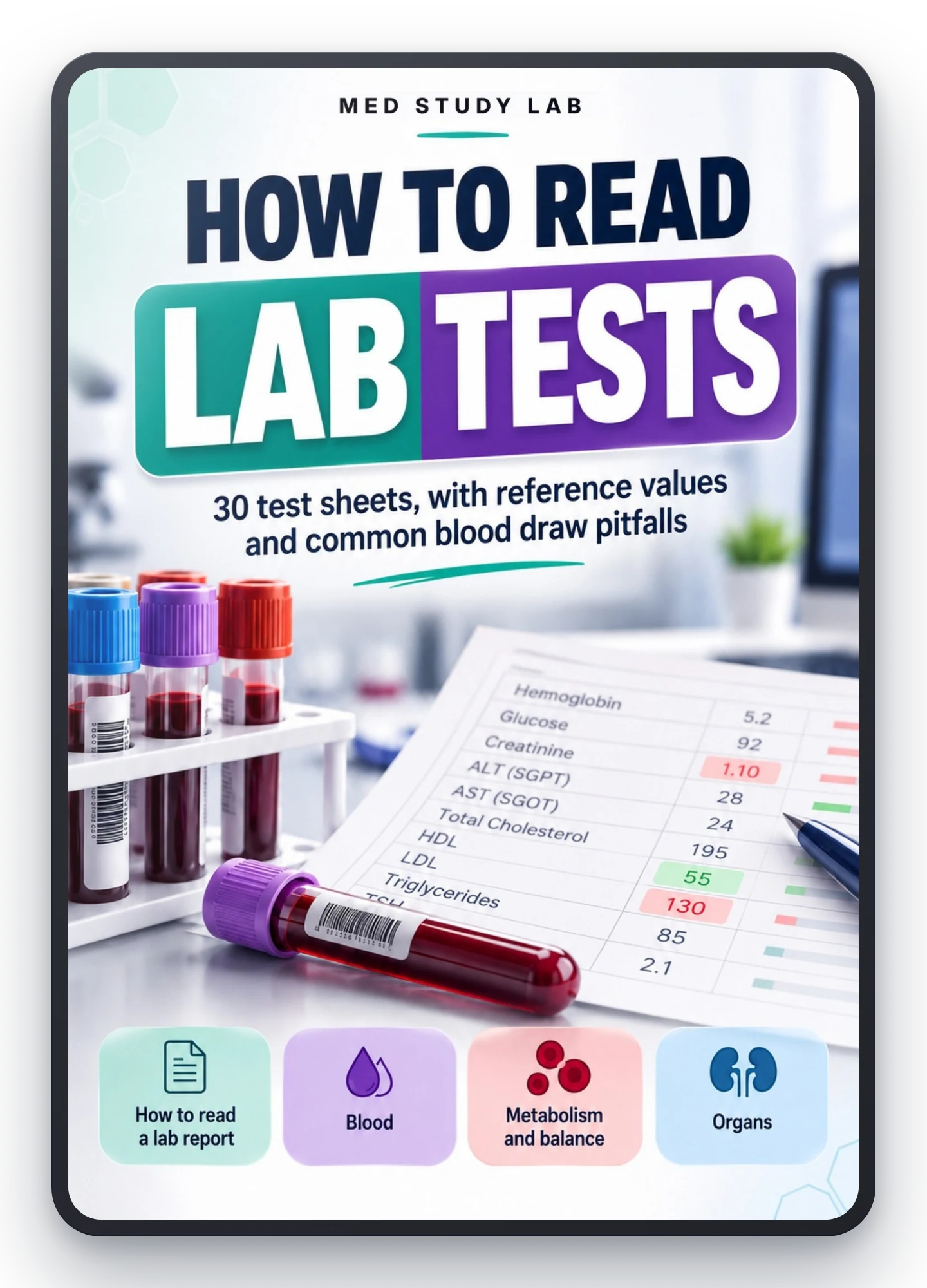
BONUS 2: “How to Read Lab Tests”
Standalone value: $17
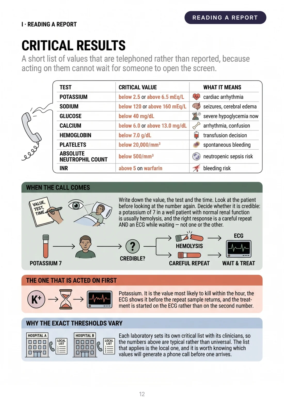
74 pages to read a lab report: potassium, calcium, troponin and the CBC, with the critical values and what throws them off.
Read it next to Part VII: a peaked T wave and a high potassium are the same emergency seen from two sides.
Reading EKGs Made Visual + 2 Bonus Books
- Reading EKGs Made Visual: 68 illustrated pages$55
- Bonus 1: Emergency Medications$19
- Bonus 2: How to Read Lab Tests$17
- High-resolution printable PDFsIncluded
Total value: $91
Today only:
$27
YOU SAVE $64
Secure purchase
Encrypted payment
30-day guarantee
All 3 books arrive by email right after purchase. One-time payment, no installments or hidden fees.
Is This Material Right for You?

Nursing or medical student
EKGs are on the exam and in clinicals, and textbooks explain them as if you already knew. You need to start from the paper and get to MI without skipping a step.

Floor, telemetry or ER nurse
The monitor alarms, the tracing changes, and you're the first to see it. You want to know in seconds whether it's artifact or you need to call right away.

New grad or on nights
On nights or on the floor, you often read the EKG alone. You need a safe method and a quick review of blocks, ischemia and electrolytes.
Who's Behind Med Study Lab

To Recap: Here's Everything You Get
- Reading EKGs Made Visual: 68 illustrated pages in 8 parts, from the EKG paper to MI and electrolytes, on real tracings
- Bonus 1: “Emergency Medications”, illustrated handbook, 82 pages (value $19)
- Bonus 2: “How to Read Lab Tests”, 74 pages (value $17)
- Delivery: all 3 books by email right after purchase
- 30-day guarantee, no questions asked: try it risk-free
- High-resolution PDFs: print them, annotate them, take them to clinicals
$91
$27
“Risk-Free Trial” Guarantee: 30 Days
You have 30 full days to try Reading EKGs Made Visual and the two bonus books.
If in that time you feel the material didn't help you, wasn't useful or simply wasn't what you expected, I'll refund 100% of your purchase.
No questions. No fine print. No hassle.
The risk is ZERO. The decision is yours.
Frequently Asked Questions
Right after purchase you get an automatic email with all three books: Reading EKGs Made Visual and the two bonus books, Emergency Medications and How to Read Lab Tests. They are high-resolution PDFs, ready to download, print or read on any device.
One-time payment of $27. No installments, no recurring charges, no surprises. You pay once and the material is yours forever.
No, you start from zero: Part I explains the paper, the leads and the waves before any arrhythmia. If you already read EKGs, you get the 8-step method and the chapters on MI and electrolytes.
Real. They come from PTB-XL, a public collection of 21,799 12-lead EKGs from 18,869 patients, read by cardiologists and published on PhysioNet. A drawn tracing is always clean; the ones you meet on the unit are not, and those are the ones to train your eye on.
No. It is study material to understand and recognize, not a clinical protocol. With a real patient you follow your facility's procedures and, when in doubt, ask: the book helps you get there prepared.
Yes, for personal use and without limits. The PDFs are high resolution, made to print, annotate and take to clinicals or the floor.
One Last Thing Before You Decide...
The EKG doesn't have to be the part you avoid.
You don't have to memorize fifty patterns without understanding why they look that way.
And you definitely don't have to find out you can't read one right when it matters.
There's an easier way. And it's one click away.
Eight steps, real tracings and two bonus books for acute care.
Now it's your turn.
- One-time payment: $27
- Guarantee: 30 days, no questions asked
- Delivery: all 3 books right after purchase
The books arrive by email within minutes